Drawing Of Protein
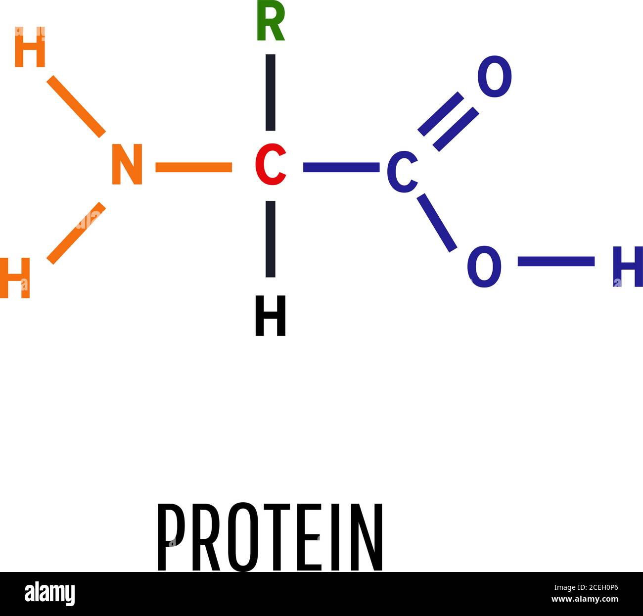
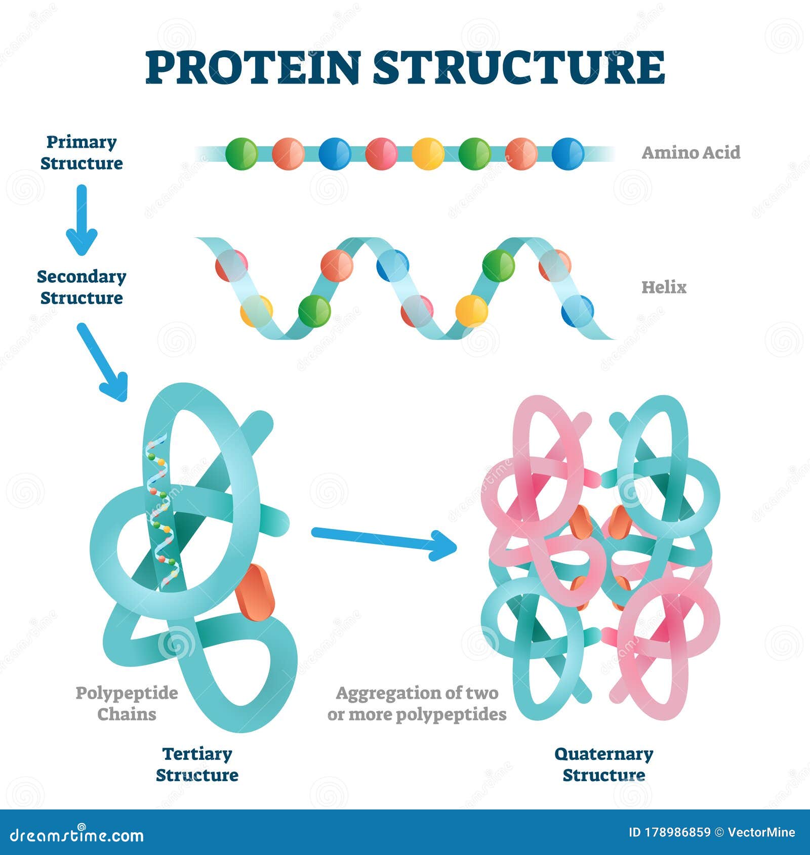
Drawing Of Protein - Primary, secondary, tertiary, and quaternary. Proteins, nucleic acids, and lipid membranes are shown; These illustrations are free for use under. Small molecules, ions, and water are omitted for clarity. A single protein molecule may contain one or more of the protein structure types: To understand how a protein gets its final shape or conformation, we need to understand the four levels of protein structure: Goodsell integrate information from structural biology, microscopy and biophysics to simulate detailed views of the molecular structure of living cells. Nearly every protein in humans is phosphorylated during its cellular lifetime to regulate cellular processes but these modifications are hard to make and study. Web the watercolor paintings of david s. Proteins consist of long polypeptide chains and we know how to determine the sequence of the amino acids of these chains. However, if you denature proteins (e.g. Web most proteins have one highly stable tertiary structure, which is often organized around a core region of hydrophobic residues. A protein is one or more polymers of monomers called amino acids. Uniprot provides multiple topological conformations for. Sketch/draw the synthesis of a hypothetical protein fragment within a cell. Web in this article we will discuss about the various structures of proteins. The search for resulted in multiple hits. Primary, secondary, tertiary, and quaternary. Web protein condensate atlas from predictive models of heteromolecular condensate composition. Web the four levels of protein structure are distinguished from one another by the degree of complexity in the polypeptide chain. Enter the pdb code in the search box and press the go button. They act as enzymes, structural support, hormones, and a whole host of other functional molecules. Primary, secondary, tertiary, and quaternary. Web most proteins have one highly stable tertiary structure, which is often organized around a core region of hydrophobic residues. Web a schematic drawing summarizes the overall. Primary, secondary, tertiary, and quaternary. Web a schematic drawing summarizes the overall features of a structure in a quickly graspable and relatively memorable form. Include the following labeled elements: Click a structure image to access its record page. Small molecules, ions, and water are omitted for clarity. Goodsell integrate information from structural biology, microscopy and biophysics to simulate detailed views of the molecular structure of living cells. Primary, secondary, tertiary, and quaternary. Small molecules, ions, and water are omitted for clarity. However, if you denature proteins (e.g. Depending on what they are interested in looking at they will pick different ways to draw and display the protein. However, if you denature proteins (e.g. Web protein condensate atlas from predictive models of heteromolecular condensate composition. This constitutes the primary structure of proteins. Depending on what they are interested in looking at they will pick different ways to draw and display the protein. Small molecules, ions, and water are omitted for clarity. Web for this journal page you are making an original labeled drawing of protein synthesis. Click a structure image to access its record page. Nearly every protein in humans is phosphorylated during its cellular lifetime to regulate cellular processes but these modifications are hard to make and study. However, for drawing the structures of proteins, we usually twist it. Web. Proteins, nucleic acids, and lipid membranes are shown; Depending on what they are interested in looking at they will pick different ways to draw and display the protein. Nearly every protein in humans is phosphorylated during its cellular lifetime to regulate cellular processes but these modifications are hard to make and study. Navigate through molecular structures, analyze interactions, and gain. Enter the pdb code in the search box and press the go button. Primary, secondary, tertiary, and quaternary structure. This could be a family portrait, a picture of a loved one, a pet, a scenic landscape, or any image that holds special. Web most proteins have one highly stable tertiary structure, which is often organized around a core region of. A single protein molecule may contain one or more of the protein structure types: Proteins consist of long polypeptide chains and we know how to determine the sequence of the amino acids of these chains. Web choose from drawing of protein foods stock illustrations from istock. Web partnering with addgene, a nonprofit plasmid repository, gce4all offers a permaphos kit for. Web most proteins have one highly stable tertiary structure, which is often organized around a core region of hydrophobic residues. This could be a family portrait, a picture of a loved one, a pet, a scenic landscape, or any image that holds special. I try to depict the overall shape of the molecule by drawing the structures as simple shapes.. However, if you denature proteins (e.g. I try to depict the overall shape of the molecule by drawing the structures as simple shapes. Primary, secondary, tertiary, and quaternary. To learn how interactions between amino acids cause a protein to fold into its mature shape, i highly recommend the video on orders of protein structure. Depending on what they are interested. They act as enzymes, structural support, hormones, and a whole host of other functional molecules. Nearly every protein in humans is phosphorylated during its cellular lifetime to regulate cellular processes but these modifications are hard to make and study. Include the following labeled elements: Web partnering with addgene, a nonprofit plasmid repository, gce4all offers a permaphos kit for academics and nonprofit organizations, make phosphorylated proteins of their choosing. I try to depict the overall shape of the molecule by drawing the structures as simple shapes. Web as we mentioned in the last article on proteins and amino acids, the shape of a protein is very important to its function. This constitutes the primary structure of proteins. 1b8g) go to the structure home page. Sketch/draw the synthesis of a hypothetical protein fragment within a cell. Web partnering with addgene, a nonprofit plasmid repository, gce4all offers a permaphos kit for academics and nonprofit organizations, make phosphorylated proteins of their choosing. Primary, secondary, tertiary, and quaternary structure. Web most proteins have one highly stable tertiary structure, which is often organized around a core region of hydrophobic residues. Navigate through molecular structures, analyze interactions, and gain insights into protein functionality. Web a schematic drawing summarizes the overall features of a structure in a quickly graspable and relatively memorable form. A single protein molecule may contain one or more of the protein structure types: Web in this article we will discuss about the various structures of proteins.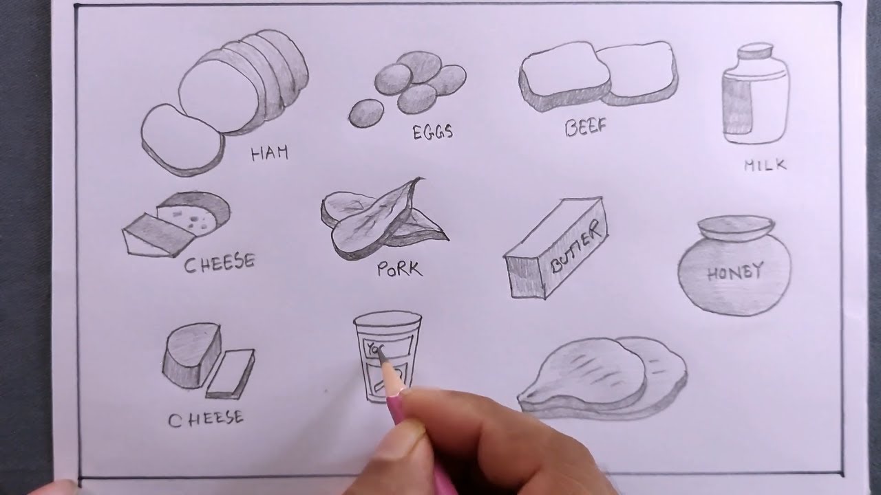
how to draw protein foods/protein foods drawing YouTube
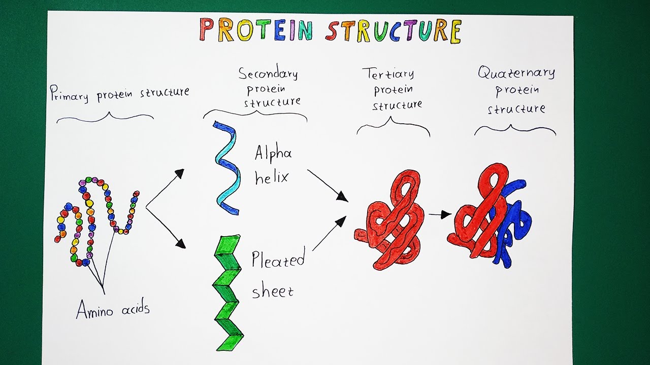
Protein Structure made EASY YouTube

How to draw Protein Foods YouTube
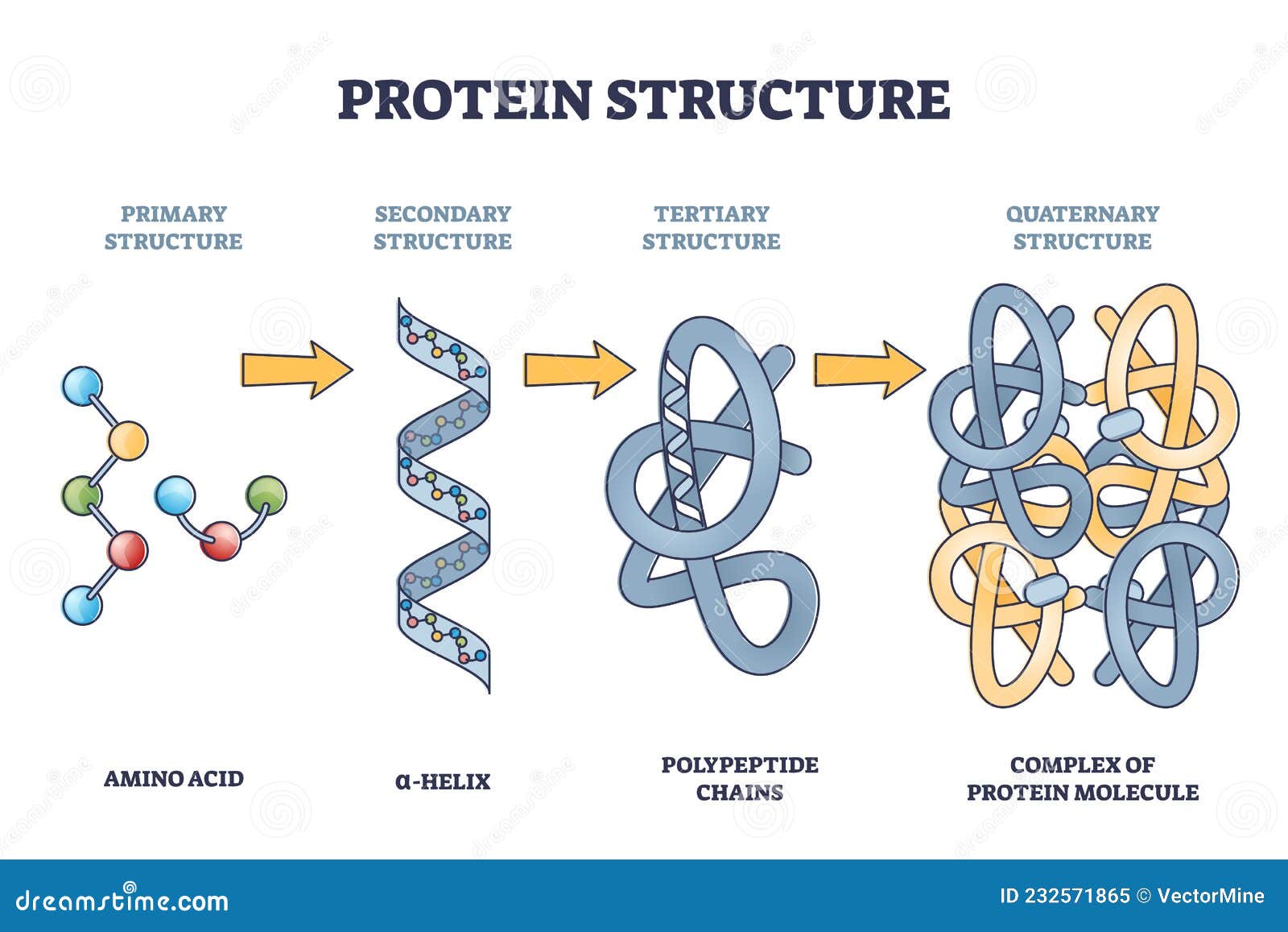
Protein Structure Levels from Amino Acid To Complex Molecule Outline
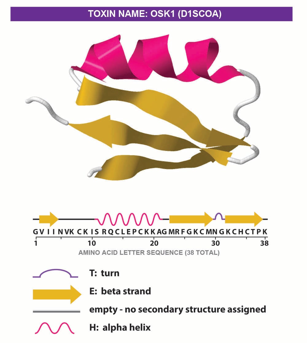
Protein Illustrations and Visualization Ask A Biologist

MCAT Biology & Biochemistry Glossary Protein Structure Class 1
Protein. Structural chemical formula and molecular model. General

Protein Foods In Pencil Colour Sketch Simple Style Stock Illustration

how to draw protein food/sources of protein food/protein food drawing
Protein Structure Vector Illustration. Labeled Amino Acid Chain
Web For This Journal Page You Are Making An Original Labeled Drawing Of Protein Synthesis.
To Understand How A Protein Gets Its Final Shape Or Conformation, We Need To Understand The Four Levels Of Protein Structure:
It Follows That There Are Two Crucial And Nontrivial Tasks Of A Schematic Drawing.
For Example, One Should Try To Draw A.
Related Post: